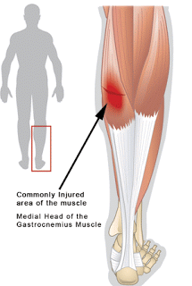
I have been treating calf injuries in runners for 10 years. I thought I had seen it all, in addition to the usual suspects like Achilles tendonitis and tendonosis I have successfully treated ruptured Achilles tendons, Soleus tendonosis and Haglund’s Deformities (pump bumps) the size of walnuts. I recently had a humbling experience that was also an “Ah Ha” moment. A patient had presented with a long history of bilateral calf pain that would come and go. He stated that after he turned forty years old he consistently started having random but severe pain in his calf(s) while running. He explained that he would be running along and all of a sudden either one or the other of his calves would begin to tighten up and would feel as if it was going to “snap”. Of course this sensation of wanting to tear would cause him to stop running and rest between a few days to a few weeks. This person was quite frustrated because this had been going on for 16 years and he had sought treatment from every kind of doctor and therapist there was without any change in his condition! Oddly, he had never had an MRI. We decided that we would get the MRI before starting any kind of treatment. I explained that I would be looking for either entrapment of his Popliteal artery which may explain his condition or the existence of a Plantaris muscle which also could account for his long standing symptoms. If this patient had presented to our clinic and this had been the first time his symptoms had appeared we would have diagnosed and treated him with a simple Gastrocnemius strain but because of the duration (16 years) and seemingly random occurrence of his symptoms I began to think about more obscure diagnosis but that still would make sense hence the Popliteal Artery entrapment and Plantaris concepts. Interestingly this individual had also been the second person in a year to describe almost the exact set of symptoms. This also triggered me to start extensively researching the various possibilities.
When the MRI came back I was not surprised. It showed exactly what the last patients MRI showed….”scarring of the medial Gastrocnemius tendon along the junction with the Plantaris tendon” with everything else showing as normal. This description by the radiologist is 100% consistent with the patient’s description of his injury. I believe that over time the Plantaris tendon has been slowly tearing and separating from its confluence with the Gastroc tendon! After exhaustive research and reading I finally found information that would lead me to believe this is exactly what is happening. The problem is that most of the people that get “Tennis Leg” rupture their Plantaris tendon instead of stopping prior to the rupture! So now it made sense. As the person is running the Gastrocnemius and Plantaris begin to tear or separate. This triggers a myospasm (http://medical-dictionary.thefreedictionary.com/myospasm) also known as muscle splinting. The muscle, in order to avoid further tearing violently contracts or seizes which feels to the patient as if they have been shot or punched in the calf. Of course this level of pain forces them to stop running immediately and “flat-foot” it back to their car or home. It also makes sense then why they have seen so many doctors and therapists without resolution. For example, they have the condition flare up so they go to the doctor who tells them to Rest, Ice, Compress and Elevate it which they do. The injury appears to get better so they start running again until BAM, it happens again so they go to another doctor having no faith in the last one. The next doctor tells them to rest and stretch which they do. Their leg feels better again so they start running until BAM there it goes again! This time they request physical therapy. After months of strengthening their calf they are cleared to run again until BAM the calf seizes again. Imagine your frustration having been a runner your entire life and now you can not consistently run without this injury stopping you dead in your tracks. You’ve tried rest, ice, different shoes, stretching, strengthening, rolling it, compression socks….you have probably even thought of having a priest perform an exorcism, but you have never had an MRI! To me that is simply careless and reckless.
Now the million dollar question is, how do you fix this condition? We are going to begin by injecting the scar tissue with Platelet Rich Plasma. After allowing the platelets to begin working on the scar tissue for roughly 1 week we are going to initiate high frequency, continuous ultrasound therapy followed by a muscle technique to help gradually break up the scar tissue. During the repair stage of the healing process we are going to begin Altered Gravity (https://www.sdri.net/services/alterg-anti-gravity-treadmill/) treadmill running at 60% of the patient’s body weight which should not generate enough force to reinjure his Gastrocnemius-Plantaris complex. Every two weeks as his muscle undergo natural and specific strengthening we will increase his body weight by roughly 10%. We believe that by gradually stressing his calves during the tissue building phase we will take advantage of Wolfe’s Law (http://medical-dictionary.thefreedictionary.com/Wolff’s+law) resulting in new and stronger calf muscle tissue being formed. After the patient progresses through the repair stage into the tissue remodeling stage we will begin having them perform an aggressive stretching routine at home in addition to continuing to progress towards 100% body weight on the AlterG. This protocol is experimental but is based on sound physiological principles and we believe are these patients best hope for full recovery and return to running.
If you have an injury and require further information email info@sdri.net or to schedule an appointment call 858-268-8525
The medical information on this site is provided as an information resource only, and is not to be used or relied on for any diagnostic or treatment purposes. This information is not intended to be patient education, does not create any patient-physician relationship, and should not be used as a substitute for professional diagnosis and treatment.


Elliptical machine did it.
After over 8 years of using the elliptical trainer, starting out at over 260lbs and working down to 180lbs, After losing all that weight, I began to try running again. The pain hit after less than a lap. Not just pain, but the kind described above.
I found that my elliptical trainer workouts suffered also and put it together. While ellipticals are easier on the knees, its hell on the plantaris tendon. I slowly nursed it with rest and ice and was able to run further and faster over a couple of years.
When doing a 7k it hit very hard. I tried running through the pain, but my calf eventually would not work. The bruising and pain are consistent with your article.
Afterwards, it was impossible to run or use the elliptical and I eventually gave up. It still aches after 5 years.
This treatment gives me hope.